endoscopic forceps laparoscopic bipolar forceps
The advent of bipolar coagulation as early as 1940. The difference between it and unipolar coagulation is that the ineffective electrode contacting the patient’s buttocks is eliminated, and the two electrodes are connected to the two leaves of a pair of tweezers. The blades are insulated. In application, the current only passes through the tissue between the two tips of the tweezers, so the required power is greatly reduced, generally only 1/4 to 1/3 of unipolar coagulation. With the development of electrosurgery technology, bipolar coagulation has become indispensable in laparoscopic surgery. Just like making eggs in egg-ham sandwiches, bipolar coagulation forceps are an important part of laparoscopic surgical instruments. Bipolar electrocoagulation is an electronic radio frequency current generator. The bipolar is in good contact with the tissue. The current passes between the bipolar poles, and the deep coagulation spreads radially, causing the blood vessels between the two ends of the bipolar to be dehydrated and coagulated. Relevant tissues are degenerate, and no obvious arc is formed.
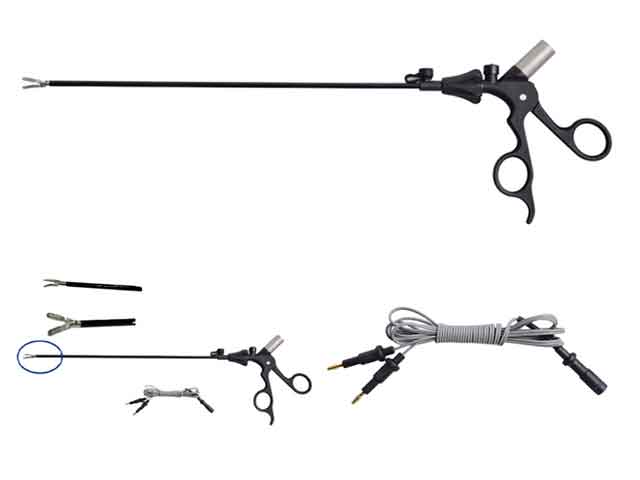
Since a loop is formed between the jaws of the bipolar forceps, there is no need for a negative plate. Bipolar forceps basically have no cutting function, mainly blood coagulation function, the coagulation speed is slow, but the hemostatic effect is reliable, because its range of action is limited to the two ends of the forceps, so the degree of damage and impact on adjacent tissues is very small. It has minimal impact on surrounding tissues. Bipolar coagulation is more accurate than unipolar coagulation. It does not need to use a negative plate, and a loop is formed between the two poles. The discharge area is very accurate, and the secondary damage is much smaller than that of unipolar. It is more conducive to hemostasis and tissue separation. When using bipolar hemostasis, try to keep the surgical field relatively dry.
Steps
1. Turn on the power, connect the pedals, and place them under the feet of the surgeon.
2. Power-on self-check, set the output power according to the requirements of the operation and the surgeon.
3. Connect the bipolar coagulation cord plug.
4. After bipolar clamp the tissue or bleeding point, step on the pedal to stop the bleeding, and then release the pedal.
5. After using, turn off the switch of the host first, and then dial the power plug.
skills
1. Commonly used precautions for bipolar
1. Choose a suitable bipolar clamp and output power 30-50W. Choose according to the nature of surgery and tissue.
2. Keep the tissue tension-free during use; keep the surgical field clean; avoid high temperature affecting the surrounding important tissues and structures; reduce the adhesion of tissue eschar and electrocoagulation forceps.
3. Each time of coagulation is within 3 seconds, and can be repeated many times until the coagulation effect is achieved. Intermittent electrocoagulation is more effective than continuous electrocoagulation to prevent the eschar of the forceps tip and tissue.
4. Remove the eschar on the bipolar forceps in time: wipe the eschar with wet gauze or a special non-damage cloth.
5. The bipolar clamp ends should be kept at a certain distance, and they should not touch each other to form a current short circuit. Loss of coagulation.
6. When coagulating near important organizational structures, the coagulation output should be as small as possible and the time should be short.
7. The bipolar forceps after use have a temperature. Avoid using it as a separate forceps, such as throwing an intestine tube, so as to avoid electric heating damage. Electric heating has different electric heat radiation conduction in different tissues, and urinary system/intestinal injuries are common.
Electrocoagulation perfect:
After ⑴ is electrocoagulated, the color of the blood vessel first changes from purple to white, and then to brownish yellow; the tube wall still maintains a certain degree of flexibility.
⑵ The blood vessel shrinks, the diameter of the blood vessel is obviously reduced, about half the diameter of the original blood vessel; the length of the blood vessel electrocoagulation is 2-4 times its diameter.
When the coagulation is completed, the tip of the forceps does not adhere to the blood vessel wall.
⑷ General external forces such as stretching, aspiration or blood pressure will not cause bleeding.
Excessive coagulation:
⑴ The color of blood vessel changes from brownish yellow to charred black, and the tube wall is hard and brittle.
⑵ The blood vessels shrank severely, and the diameter was less than 1/3 of the original.
⑶ The tip of the forceps may stick to the wall of the tube.
⑷ can not withstand the slight influence of external force, it is easy to break and bleed.
Insufficient coagulation:
⑴ The color of blood vessel changes from purple to white only.
⑵ The blood vessel shrinks very little, the diameter of the blood vessel is not significantly reduced or becomes smaller and then expands immediately; or the length of the blood vessel electrocoagulation is not enough.
⑶ can not withstand the slight influence of external force and bleed again.
3, bipolar coagulation hemostasis method
The method we adopted can be summarized into six essentials:
⑴ Choose a wider forceps tip (most commonly 5mm) and a lower coagulation output to avoid excessive coagulation or adhesion of the forceps tip to the vessel wall.
⑵ Intermittent Coagulation: It is not prone to excessive electrocoagulation or adhesion of the clamp tip to the vessel wall. Coagulation is performed for about 3 seconds each time, and repeated several times until it reaches the perfect standard of electrocoagulation.
⑶ Migrating incremental electrocoagulation method: For larger arteries, conduct electrocoagulation gradually from the proximal end of the blood vessel to the distal end, and gradually increase the number of intermittent electrocoagulation until the surface of the distal blood vessel is blackened, and cut at the darkened place. Blood vessels.
⑷ The length of the vascular cauterization zone should be 2-4 times its diameter, and try to cut it distally. After electrocoagulation, the tissue can be moistened with normal saline to avoid excessive electrocoagulation or electrothermal damage. Because of the thin wall of the venous blood vessel, it is easy to achieve satisfactory cauterization under conventional electrocoagulation. On the other hand, if the electrocoagulation conditions are not well controlled, it is prone to puncture of the vessel wall, adhesion and tearing, etc.